
Up to 24 million Americans have been diagnosed with at least one autoimmune disease, and 63% of them are women. That is likely a significant undercount, since many people with autoimmune thyroid disease are labeled “hypothyroid” and never tested for antibodies.
Autoimmune numbers are climbing. Positive antinuclear antibody (ANA) tests, a sign the immune system is reacting against the body’s own tissue, almost doubled from 1988–1991 to 2011–2012. The steepest rise was in teenagers. Genes don’t change that fast, and considerable evidence points to our environment.
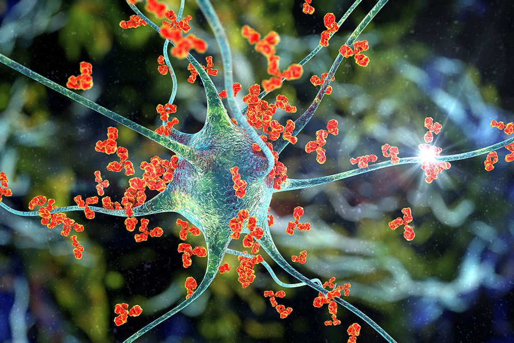
If you have one autoimmune condition, you are at a greater risk for developing another. This tracks since underlying causes of autoimmune conditions are rarely explored. Whatever contributed to one disease can lead to another.
More than 100 autoimmune conditions exist. In this article, I focus on three common autoimmune conditions strongly associated with chronic infections and environmental toxins that can drive them.
- Hashimoto’s disease (autoimmune thyroiditis): the immune system attacks the thyroid.
- Multiple sclerosis (MS): the immune system attacks the myelin that insulates nerves in the brain and spinal cord.
- Rheumatoid arthritis (RA): the immune system attacks the joints.
How Infections and Toxins Turn the Immune System Against the Body
Infections
The best-known mechanism by which infections cause autoimmune conditions is molecular mimicry. In molecular mimicry, an infection’s protein resembles a human one, so antibodies directed at the infection also attack our own tissue. Rheumatic fever after strep throat is the classic example.
Infections also damage tissue and expose proteins the immune system never learned to tolerate, and inflammation can wake dormant self-reactive cells. Some viruses, like Epstein-Barr virus (EBV), hide in immune cells for life and periodically reactivate, causing a low-level immune activation.
Environmental Toxins
Toxins damage cells and chemically alter our proteins so they no longer look like our own tissue. Mercury is a well-studied example. Genetically susceptible mice given low doses of mercury develop autoantibodies within about two weeks.1 Mercury inflames the tissue, activates immune cells, and triggers antinuclear antibodies.
People often believe they develop a disease because of genetic predisposition. As the saying goes, “genes load the gun, but the environment pulls the trigger.”
Diagnosing it early with ANA
A positive ANA (antinuclear antibody) test means the immune system makes antibodies against the nucleus of our own cells. These antibodies can appear years before an autoimmune disease begins, so it can be thought of as an autoimmune screening test and should be included in annual lab testing. This is when finding and removing the underlying drivers is critical. When the underlying causes are effectively treated, I see positive ANA levels normalize.
Hashimoto’s disease
Hashimoto’s is the most common cause of hypothyroidism in the United States. About 11.6 million Americans have autoimmune thyroid disease, and 8.8 million are women. About 4 million additional Americans have undiagnosed autoimmune thyroid disease. The most common age range at diagnosis is 30 to 60, but it can affect people of any age. Hashimoto’s disease is associated with hypothyroidism (underactive), whereas Graves’ disease is related to hyperthyroidism (overactive).
Mercury and the thyroid
Mercury is one of the main toxins I look for in Hashimoto’s disease. For most people, mercury exposure comes from eating large fish such as ahi, tuna, swordfish, shark, and king mackerel. Other sources include dental amalgam fillings. Mercury is stored in human tissues, so a person could have eaten a lot of sushi or canned tuna in their teens or 20s and still have elevated levels in their 40s or 50s.
The thyroid is like a canary in a coal mine and is easily damaged by environmental toxins
Unfortunately, mercury accumulates in the thyroid. In autopsy tissue from 115 people, researchers found mercury in the thyroid hormone-producing cells of 4% of people under 30, 9% of those 30 to 59, and 38% of those over 60.2 Earlier studies found five times more thyroid mercury in people with more than 11 amalgam fillings than in those with fewer than four. Cadmium and lead often coexisted, and these metals amplify each other’s toxicity.
Mercury can harm the thyroid in several ways:
- It binds sulfur and selenium, which thyroid cells are rich in, likely explaining why it lodges there.
- It causes oxidative damage and depletes glutathione in a gland that already uses hydrogen peroxide to make hormones.
- It breaks immune tolerance, inflames tissue, and exposes cell fragments that drive autoantibodies.
- It ties up selenium, which the thyroid needs to activate hormones and protect itself. Low selenium alone is linked to thyroid autoimmunity.
In the 2007–2008 NHANES survey, women 20 and older in the highest fifth of blood mercury were more likely to test positive for thyroglobulin antibodies. Mercury isn’t the only cause of Hashimoto’s, but anyone with hypothyroidism should be properly tested for mercury burden with a 6-hour urine provocation test.
Endocrine-disrupting chemicals
Many common environmental toxins also disrupt the thyroid. A 2024 Review3 of exposures from fetal life to adulthood describes these:
- Perchlorate – fireworks, some fertilizers, rocket fuel found in water and produce; blocks iodine from entering the thyroid.
- Bisphenols – BPA and “BPA-free” substitutes like BPS, in plastics, can linings, and receipts, block iodine uptake and thyroid hormone receptors. Exposure in pregnancy is linked to higher TSH (hypothyroid) in newborns.
- Phthalates, found in soft plastics, fragrances, and personal care products, block iodine uptake and T3 binding and are linked to altered thyroid levels in children and teens.
- PCBs – banned but persistent in fatty fish, meat, and dairy and related dioxins are associated with lower thyroid hormone and more hypothyroidism.
- PFAS – “forever chemicals” in nonstick pans, stain-resistant fabric, food packaging, and water are linked to lower T4 in infants and faster hormone breakdown.
These chemicals mainly disrupt hormone production rather than trigger antibodies. But a thyroid already under autoimmune attack has less room to compensate, and people with thyroid antibodies may be more vulnerable. Exposure before birth is the biggest concern, so testing for environmental toxins should be standard of care before conception.
Multiple sclerosis
Nearly 1 million American adults have MS, more than double previous estimates. It is usually diagnosed between ages 20 and 50, and women are affected about three times more often than men. Rates are highest among whites and those living in northern latitudes.
Epstein-Barr virus and other infections
The strongest evidence that an infection can cause autoimmune disease comes from MS and Epstein-Barr virus. Researchers tracked more than 10 million young adults in the U.S. military for up to 20 years using stored blood samples. EBV infection raised the risk of getting multiple sclerosis 32-fold,4 and a marker of nerve damage increased only after infection. No other virus tested showed this pattern. The authors concluded EBV is the leading cause of MS.
One possible mechanism associating EBV with MS is molecular mimicry. Part of the EBV protein EBNA1 (Epstein-Barr nuclear antigen-1) resembles a protein in the brain and spinal cord called GlialCAM, so EBV antibodies can cross-react and attack human tissue.
Other infections add to the risk. In a study of over 8,000 MS patients, antibodies to human herpesvirus 6A (HHV-6A) were linked to higher MS risk, especially in conjunction with high EBV antibody levels.5 There is also evidence that Lyme disease can contribute to MS,6 and I have seen the association in my practice.
Lyme can cause nerve inflammation and brain lesions that mimic MS, which may actually be an MS-like presentation of Lyme rather than MS.
If a person’s health history includes a tick bite or activity that increased tick-bite risk, I always rule out tickborne infections with accurate lab testing.
Mercury and Multiple Sclerosis
Mercury is a potent neurotoxin, and studies of people with mercury toxicity have reported symptoms of numbness, tremor, vision loss, and trouble walking, symptoms that are consistent with MS.
One study compared people with relapsing-remitting MS to matched controls.7 The average serum mercury level was almost twice as high in the MS group compared to controls, and people with MS were almost 2.5 times more likely to have abnormally high mercury levels. In lab studies, even low doses of mercury kill oligodendrocytes, the nervous system cells that make myelin and are damaged in MS.
Because mercury damages the same tissue as MS, I believe it deserves to be properly tested with a 6-hour provoked urine toxic metal test in anyone with MS. I’ve written about a case of MS that was caused by mold and mercury toxicity.
Mold and fungal toxins in Multiple Sclerosis
Mold is an underappreciated piece of the multiple sclerosis puzzle. Researchers proposed that toxins from molds such as Aspergillus, Candida, Fusarium, and Penicillium may drive the myelin damage characteristic of MS.8 These fungi can hide from the immune system while releasing toxins that cross the blood-brain barrier and kill astrocytes and oligodendrocytes, the cells that protect myelin.
In animal studies, gliotoxin, produced by the mold Aspergillus and the yeast Candida, destroys these cells. Spinal fluid from MS patients contains a factor that kills the same cells, which spinal fluid from other neurological diseases does not. A protein with biochemical characteristics identical to those in gliotoxin and is toxic to glial cells was found in the urine from 32 of 35 MS patients. In addition, fumonisin B1, from Fusarium, disrupts the fats that make up myelin.
In patients with MS and a history of mold exposure, I always test urine mycotoxins
Other environmental toxins in MS
Other exposures linked to MS include organic solvents, air pollution and particulate matter, and pesticides, PCBs, and PFAS. Smoking delivers many of these toxins at once and is an established MS risk factor.
Rheumatoid arthritis
Lyme disease and Bartonella
Tickborne infections can cause joint disease that mimics rheumatic illness, and they are rarely tested for. One study tested blood and joint fluid from 110 patients with swollen joints in a rheumatology practice.9 Over a quarter (27%) had antibodies to Borrelia burgdorferi (Lyme disease) and 24% had antibodies to Bartonella henselae or B. quintana. Lyme antibodies were found more often in joint fluid than in blood (27% vs. 15%), suggesting blood tests alone can miss Lyme in the joints.
Bartonella can also mimic rheumatologic disease. A case report was published of A 31-year-old veterinarian who developed tendon inflammation, joint subluxations, and hypermobility severe enough to be diagnosed as Ehlers-Danlos syndrome. Her rheumatoid factor and Lyme tests were negative.10 Specialized testing found Bartonella koehlerae and B. henselae in her blood, and her joint pain and hypermobility resolved with long-term antibiotics. In patients with arthritis and a history of tick bites, flea exposure, or animal contact, I test for these infections with specialized laboratories.
Heavy Metals
Smoking roughly doubles RA risk, and cigarette smoke is most people’s main source of cadmium, one reason researchers suspect metals are part of that link. Cadmium and lead accumulate for decades, generate oxidative stress, and alter immune responses. In animals, mercury causes the same inflammation, autoantibodies, and immune-complex deposits that damage joints in RA. Human studies of individual metals in RA remain few and inconsistent. In my experience, metals are usually one stressor among several, not the sole cause.
Other Environmental Toxicants
RA often begins in the lungs. Inhaled irritants trigger citrullination, a protein modification that produces the anti-CCP antibodies seen in most RA patients, often years before joint pain. Besides smoking, the strongest evidence is for silica dust from construction, mining, and stone countertop work, which raises risk two- to threefold. Textile dust, pesticides, solvents, and traffic pollution are also linked.
Mycoplasma
Mycoplasmas are tiny bacteria without a cell wall that can invade cells and evade the immune system. Several mycoplasma species cause chronic arthritis closely resembling RA. In one study, researchers used sensitive PCR testing to detect mycoplasma DNA in the white blood cells of people with RA and healthy controls.11 More than half of the RA patients (54%) tested positive, compared to 9% of controls. The authors concluded that systemic mycoplasma infection may be a cofactor in RA. Previous trials demonstrated that the antibiotic minocycline modestly improved RA, suggesting an infectious association. It is worth testing for mycoplasma infections in rheumatoid arthritis, especially when arthritis began after a respiratory illness.
Epstein-Barr Virus
People with RA tend to carry more EBV in their blood, and EBV has been found in the joint lining. An EBV protein, gp110, contains the same amino acid sequence as the HLA shared epitope that confers RA risk, and antibodies to citrullinated EBV proteins cross-react with the proteins targeted in RA. A poorly controlled EBV infection may help start and sustain the attack on the joints.
Looking for the Fire
Scientific research has identified chronic infections and environmental toxins as potential causes of certain autoimmune conditions. EBV raises MS risk 32-fold and is implicated in rheumatoid arthritis. Lyme, Bartonella, HHV-6, mycoplasma, and other persistent infections add to the burden. Mercury accumulates in the thyroid and nervous system and triggers autoimmunity in multiple sclerosis and autoimmune thyroiditis. Mold toxins, silica, solvents, PFAS, PCBs, bisphenols, and phthalates disrupt immune competency and can lead to autoimmunity. Genes determine who is vulnerable, but infections and toxins influence genetic expression. Unlike genes, these exposures can be identified and treated.
The body doesn’t start attacking itself without a cause. Autoimmunity is the immune system responding, often for years, to something it perceives as a threat.
This is where mainstream medicine falls short. The standard approach is to suppress the immune system with steroids, immunosuppressants, and biologics, or replace what’s been lost, like thyroid hormone. These treatments can help, but they don’t address why the immune system turned on the body. It’s like treating the smoke without looking for the fire.
In my practice, the goal is to find the fire. That means taking a careful medical history, ordering accurate lab tests for chronic infections and toxic burden, and creating an effective treatment plan so the immune system can regain competency.